Anatomy Of Upper Thigh And Hip / Hip Pain Explained Including Structures Anatomy Of The Hip And Pelvis : He also serves the communities of charleston, sc and augusta, ga.
byAdmin-
0
Anatomy Of Upper Thigh And Hip / Hip Pain Explained Including Structures Anatomy Of The Hip And Pelvis : He also serves the communities of charleston, sc and augusta, ga.. The thigh is the area between the hip and the knee joint. Foundational anatomy provides medical students with the necessary background in anatomy for success in clerkships. Iliopsoas muscle, a hip flexor muscle that attaches to the upper thigh bone. Knowing the anatomy of your hip can help you understand the source of any hip pain. Want to learn more about it?
This deep muscle begins in the low back and pelvis and connects on the inside edge of the upper femur. for detailed anatomy of pelvic bones, read anatomy of hip bone. The upper part of the thigh bone consists of the femoral head, femoral neck, and greater and lesser trochanters. The different anatomical areas of the gluteal region: Groin, inguinal region and the anterior and posterior regions of the hip and thigh.
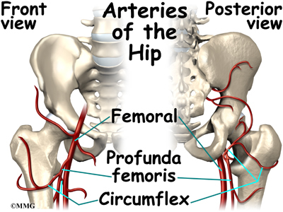
Blood Supply Of The Thigh Anatomy Orthobullets from upload.orthobullets.com While the thigh muscles will be slip into the anterior, medial and posterior groups. Upper part of the ischial tuberosity insertion: Hip flexor deep in pelvis a composite o… used to extend the hip when climbing st… The hip joint is a ball and socket joint that is the point of articulation between the head of the femur and the acetabulum of the pelvis. Hip anatomy, function and common problems. 340 anatomical structures of the hip region were labeled, accessible on anatomical parts: This arrangement gives the hip anatomy a large amount of motion needed for daily activities. The upper part of the thigh bone consists of the femoral head, femoral.
Hip anatomy, function and common problems.
This vein, as well as the deep veins. Like the forearm, the upper leg, or thigh, has a dense arrangement of many muscles. 431).—at the upper and medial part of the thigh, a little below the medial end of the inguinal ligament, is a large. B, muscles of the anterior thigh compartment. The different anatomical areas of the gluteal region: The paired hip bones are connected. for detailed anatomy of pelvic bones, read anatomy of hip bone. This deep muscle begins in the low back and pelvis and connects on the inside edge of the upper femur. The hip joint is a ball and socket joint that is the point of articulation between the head of the femur and the acetabulum of the pelvis. Hip surgeon dr guillaume dumont offers hip pain treatments in columbia, sc. This webpage presents the anatomical structures found on thigh mri. Knowing the anatomy of your hip can help you understand the source of any hip pain. Want to learn more about it?
The femur or thigh bone is one of the longest bones in the human body. Foundational anatomy provides medical students with the necessary background in anatomy for success in clerkships. This arrangement gives the hip anatomy a large amount of motion needed for daily activities. The uppermost of the medial thigh muscles is the pectineus muscle. The median cubital vein (a common site site for venepuncture) in the antecubital fossa of the arm.
Hip Anatomy Orthogate from www.eorthopod.com Want to learn more about it? Anatomy of the human body. B, muscles of the anterior thigh compartment. The following nerves serve the gluteal and. The femur or thigh bone is one of the longest bones in the human body. Medial condyle of tibia nerve supply: Atlas of human anatomy in cross section. The single bone in the thigh region is called the origin:
Groin, inguinal region and the anterior and posterior regions of the hip and thigh.
Want to learn more about it? Upper part of the ischial tuberosity insertion: 431).—at the upper and medial part of the thigh, a little below the medial end of the inguinal ligament, is a large. Anatomy of the human body. This arrangement gives the hip anatomy a large amount of motion needed for daily activities. Like the forearm, the upper leg, or thigh, has a dense arrangement of many muscles. The anatomical areas found on the upper limb can serve as key landmarks to help us find important anatomical structures such as finding one of the superficial veins: Hip flexor deep in pelvis a composite o… used to extend the hip when climbing st… Unlike the shoulder girdle, the pelvic girdle is firmly integrated into the axial skeleton: Pelvis, perineum, hip, and upper thigh. The cavity of the acetabulum faces obliquely forward, outward, and downward. Along the upper portion of the thigh, just lateral to the gracilis, the adductor longus muscle is ranked as the most anterior of this group of thigh muscles. It is part of the lower limb.
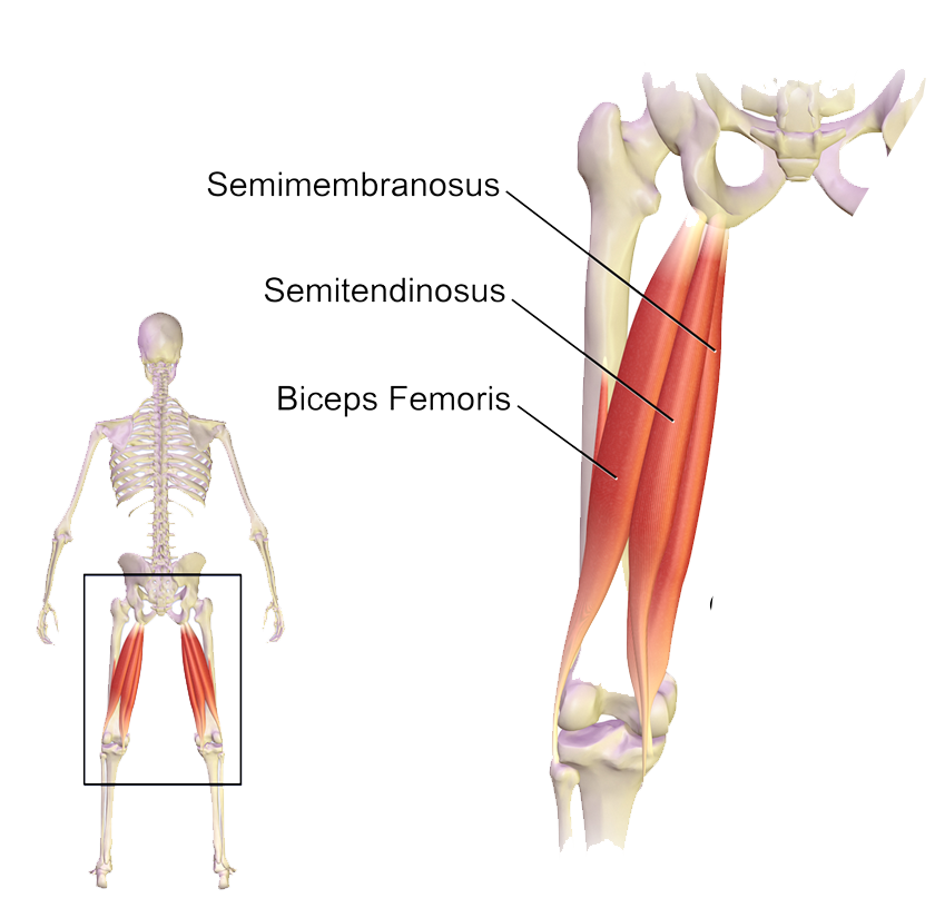
Muscles of the hips and thighs. Hip surgeon dr guillaume dumont offers hip pain treatments in columbia, sc. It functions to adduct the thigh and to flex. This webpage presents the anatomical structures found on hip mri. He also serves the communities of charleston, sc and augusta, ga.
Muscles Of The Hips And Thighs Human Anatomy And Physiology Lab Bsb 141 from s3-us-west-2.amazonaws.com Chief flexor of knee weak. Hip surgeon dr guillaume dumont offers hip pain treatments in columbia, sc. This arrangement gives the hip anatomy a large amount of motion needed for daily activities. He also serves the communities of charleston, sc and augusta, ga. A, anterior and posterior views show the hip joint ligaments. Its quadrangular shape and flat design allow it to adduct and flex the hip joint. The muscles also require a lot of blood flow, which provides oxygen and nourishment, especially when you're physically active. Jew anatomy atlases, the anatomy atlases logo, and a digital library of anatomy information are all trademarks of michael p.
During hip replacement surgery, your surgeon removes the upper part of your thigh bone, including the femoral head (ball of the hip joint) and a part the upper part of the thigh bone is then exposed, and a series of tools called broaches are introduced one at a time to prepare your thigh bone for a metal.
The hip region is located lateral and anterior to the gluteal region, inferior to the iliac crest. The uppermost of the medial thigh muscles is the pectineus muscle. The single bone in the thigh region is called the origin: This webpage presents the anatomical structures found on hip mri. The upper part of the thigh bone consists of the femoral head, femoral neck, and greater and lesser trochanters. Pelvis, perineum, hip, and upper thigh. Upper part of the ischial tuberosity insertion: The following nerves serve the gluteal and. It's restricted by contact of the thigh with all the abdomen and adduction is restricted by contact. It functions to adduct the thigh and to flex. Iliopsoas muscle, a hip flexor muscle that attaches to the upper thigh bone. He also serves the communities of charleston, sc and augusta, ga. Anatomy of the human body.
Chief flexor of knee weak upper thigh anatomy. While the thigh muscles will be slip into the anterior, medial and posterior groups.